In order to fully understand how the knee works a little information on the anatomy of the joint itself can be very helpful.
The knee is a hinge joint. Essentially this means it moves in one plane (into flexion [bending] and extension [straightening]), there also some other sliding movements that can occur. In order to keep the knee stable, there are multiple structures that hold it in place, and enable it to move smoothly. If you have an injury to the knee, pain, or difficulty with movement, it is likely that one or more of these structures may be affected. The knee joint can withstand forces of 4-6 times your body weight when loading it and doing activity. Therefore it is very important to try and keep it in good working order!
Below is a basic overview of these structures:

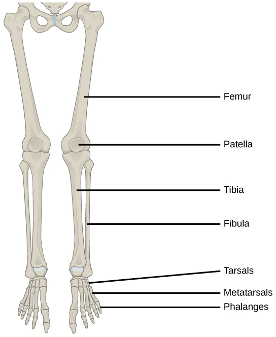
Bones
There are 4 bones that comprise the knee joint:
Femur. The longest bone in the body, and the thighbone (the bone of the upper leg).
Patella. also known as the kneecap, is approximately 3-5 cm and can vary slightly in shape. The patella plays an important role in protecting the joint underneath and is controlled primarily by the quadricep muscles. The patella slides in an ‘up and down’ motion when you bend, or straighten, your knee.
Tibia. This is the shinbone, and is the larger bone on the inside of the lower leg.
Fibula. This is the smaller bone on the outside of the lower leg.
Muscles
There are multiple muscles that help to support the knee. The two main groups are:
The quadricep muscles (thigh muscles), which attach all around the knee and onto the patella. These help to straighten your knee.
The hamstring muscles are on the back of your thigh. These help to bend your knee.
Cartilage – this is a smooth surface that covers the end of the bones. This is often confused with the meniscus which is often referred to as cartilage. The cartilage can wear away over time exposing the bone underneath.
Mensicus – this is made up of collagen fibres, and proteoglycans (energy/shock absorbers), and sits in the middle of the joint. It helps protect the joint. You can consider it a little like silicon in terms of its structure.
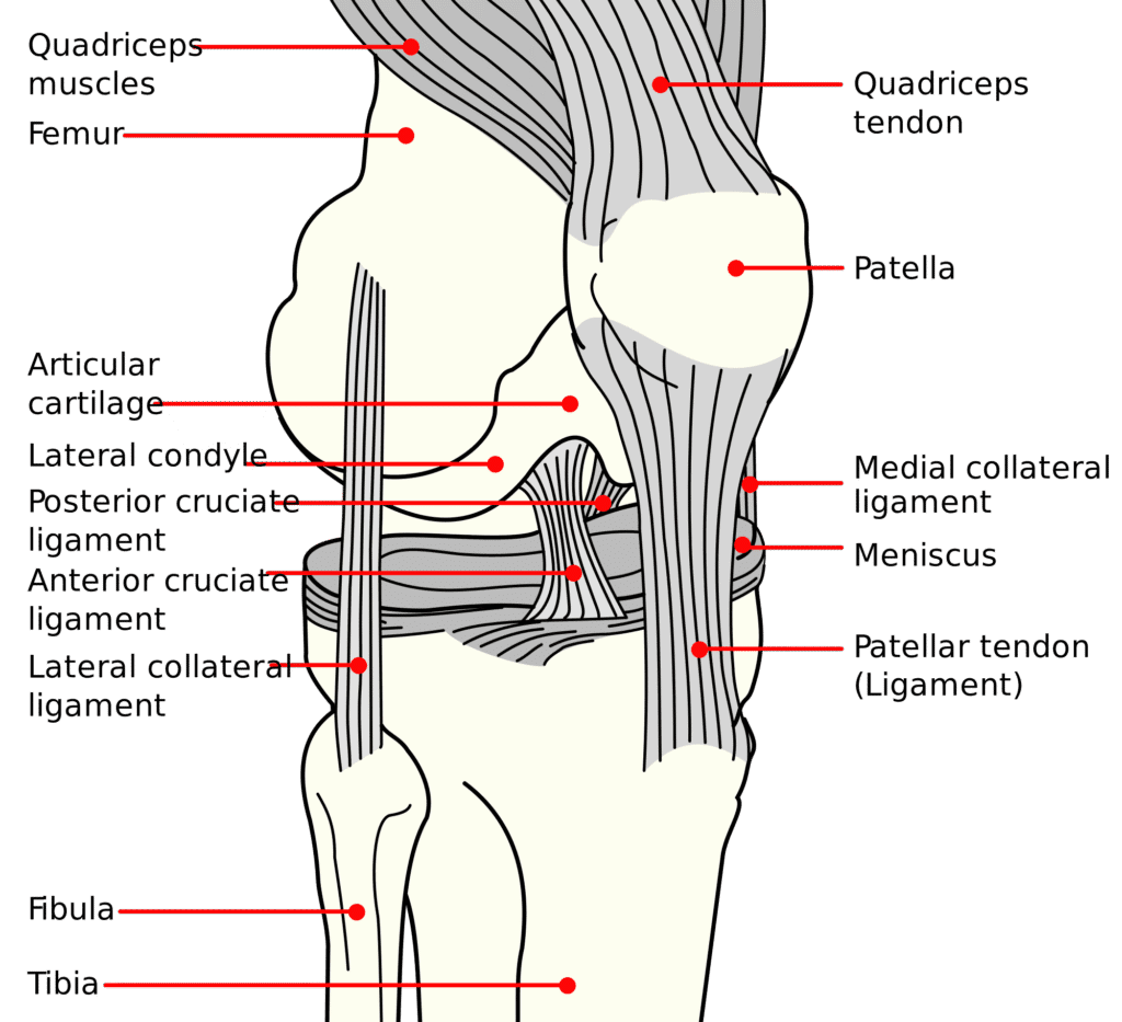
Ligaments and Tendons – there are many ligaments and tendons to support the knee in and around the joint. The four main ligaments are:
The Anterior Cruciate Ligament (ACL) – this stops your shin bone from sliding too far forwards.
The Posterior Cruciate Ligament (PCL) – this stops your shin bone from sliding too far backwards.
The Lateral Collateral Ligament (LCL) – this is on the outside of your knee, and helps stop your knee bending outwards.
The Medical Collateral Ligament (MCL) – this is on the inside of your knee, and helps stop your knee bending inwards.
The Patella Tendon goes between the kneecap and the tibia bone, and helps with the glide of the kneecap.
In addition to all of the above you also have arteries supplying the blood to the joint and nerves giving you sensation and feeling in and around the joint.
LOOK AFTER YOUR KNEES!
